Cattle, or cows, are crucial livestock globally, belonging to the species Bos taurus, and have been domesticated for over 10,000 years for diverse purposes.
These large, hoofed mammals are essential for milk, meat, and labor, exhibiting significant anatomical features that define their functionality and agricultural importance.
Understanding bovine anatomy—particularly external body parts—is vital for animal husbandry, veterinary medicine, and appreciating their biological adaptations.
Detailed anatomical diagrams, often found in PDF resources, illustrate the cow’s head, neck, torso, limbs, skin, and tail, aiding comprehensive study.

The external structure directly relates to their ruminant digestive system and behavioral cues, making observation a key aspect of animal welfare assessment.
Analyzing external features provides insights into a cow’s health, breeding status, and overall well-being, crucial for effective farm management practices.
What Defines a Cow?
Defining a cow extends beyond simple categorization; it’s rooted in biological classification and practical usage. While “cow” colloquially denotes all domestic cattle, scientifically, it specifically refers to a mature female of the species Bos taurus.
However, common language often applies the term to young cattle of either sex, and even castrated males. This broad usage highlights the animal’s significance in agriculture and daily life. Externally, a cow is characterized by a large, sturdy build, featuring a distinct head, neck, torso (or barrel), and four limbs ending in hooves.
PDF resources detailing bovine anatomy emphasize these external features, showcasing variations in coat color and patterns. The presence of a mammary system – the udder – is a defining characteristic of female cattle, crucial for milk production.
Furthermore, observing external cues like body condition and posture provides insights into a cow’s health and nutritional status. Understanding these external attributes is fundamental to proper livestock management and veterinary care.
These external characteristics are often detailed in anatomical diagrams available in PDF format.
Taxonomic Classification: Bos taurus
Bos taurus, the scientific name for the domestic cow, places it within the broader taxonomic framework of the animal kingdom. Belonging to the family Bovidae – which includes cattle, sheep, and goats – Bos taurus is an even-toed ungulate, characterized by its cloven hooves.
Externally, this classification manifests in a robust, four-legged structure adapted for grazing and locomotion. PDF anatomical guides illustrate the specific skeletal and muscular arrangements supporting this form. The species exhibits considerable variation in coat color, ranging from black and white to red and brown, often detailed in visual resources.
Detailed PDF documents showcase the external morphology, including the head with its horns (present in many breeds), the elongated neck, and the barrel-shaped torso. These external features are directly linked to the animal’s digestive system and overall physiology.
Understanding this taxonomic placement aids in comprehending the cow’s evolutionary history and its adaptation to various environments. External characteristics, as depicted in anatomical PDFs, are key identifiers within this species.
Historical Significance of Domestication
The domestication of Bos taurus, over 10,000 years ago, fundamentally altered both human societies and the cow’s own evolution. Initially valued for work and meat, selective breeding gradually emphasized traits like milk production, influencing external characteristics.
Early depictions of cattle, often found in archaeological records and illustrated in modern PDF guides, reveal changes in body size and conformation over time. Horn shapes, coat patterns, and overall musculature were intentionally modified by humans.
PDF resources detailing bovine anatomy demonstrate how domestication impacted skeletal structure, particularly in relation to load-bearing capacity for draft work. The udder, crucial for milk production, also underwent significant development, becoming a prominent external feature.
The historical interplay between humans and cattle shaped the diversity of breeds we see today, each exhibiting unique external traits. Studying these changes, as documented in anatomical PDFs, provides insight into the long-term consequences of artificial selection.
The Cow’s Head
Cows’ heads feature prominent structures like skulls, horns, eyes, ears, noses, mouths, and tongues, detailed in anatomical PDF guides.
These PDFs illustrate external features crucial for sensing, feeding, and interacting with the environment, vital for bovine survival.
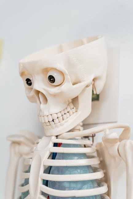
Skull Structure and Horns
The bovine skull, extensively detailed in anatomical PDF resources, is a robust structure protecting the brain and supporting facial features. These PDFs showcase the complex arrangement of cranial bones, providing a foundational understanding of head morphology.
Horns, present in many cattle breeds, are permanent cranial projections growing from the frontal bone; their size and shape vary significantly between breeds and sexes, as illustrated in detailed diagrams.
These PDF guides often depict the horn’s keratinous sheath over a bony core, explaining their function in defense, social dominance, and potentially thermoregulation. Hornless cattle, naturally or through genetic selection, are also represented.
Understanding the skull’s structure and horn development is crucial for veterinary procedures, breed identification, and assessing animal welfare, all readily available through comprehensive PDF anatomical studies.
Detailed PDFs also show the skull’s impact on the placement of sensory organs, like eyes and ears, influencing the cow’s perception of its surroundings.
Eyes and Vision
Bovine eyes, thoroughly illustrated in anatomical PDF guides, are large and positioned laterally on the head, granting a wide field of vision crucial for predator detection. These PDF resources detail the eye’s external structures, including eyelids, eyelashes, and the nictitating membrane for protection.
Cows possess nearly 360-degree panoramic vision, but with limited depth perception directly in front and behind. PDF diagrams highlight this unique visual capability, explaining its implications for handling and management.
Their dichromatic vision—perceiving primarily blues and yellows—differs from human trichromatic vision, impacting their response to colors, as shown in comparative PDF analyses.
Understanding bovine vision is vital for designing safe and efficient handling facilities, minimizing stress, and promoting animal welfare, all detailed in specialized PDF documentation.
Detailed PDFs also explain how eye placement influences their reaction to movement and shadows, impacting their behavior in pasture and confinement settings.
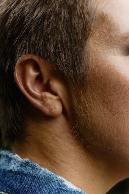
Ears and Hearing
Bovine ears, extensively depicted in anatomical PDF resources, are relatively large and mobile, capable of independent movement to pinpoint sound sources. These PDF guides showcase the external pinna’s structure, crucial for sound collection and amplification.
Cows possess a wide hearing range, particularly sensitive to frequencies between 50 Hz and 8 kHz, vital for detecting vocalizations and potential threats, as detailed in comparative PDF studies.
While their hearing isn’t as acute as some species, their ability to rotate ears allows for precise sound localization, a feature highlighted in detailed PDF anatomical charts.
Understanding bovine auditory perception is essential for minimizing stress during handling, as loud noises can induce anxiety and affect behavior, explained in welfare-focused PDFs.
PDF documentation also illustrates how ear position can indicate a cow’s emotional state, serving as a valuable behavioral cue for farmers and veterinarians.
Nose and Olfactory System
The bovine nose, thoroughly illustrated in detailed anatomical PDF guides, is a prominent feature crucial for olfaction – a cow’s highly developed sense of smell. These PDF resources demonstrate the external nasal structure and its connection to the internal olfactory receptors.
Cows rely heavily on their sense of smell for identifying food sources, detecting predators, and recognizing other individuals, as explained in ethological PDF studies.
PDF diagrams reveal the large nasal cavity and complex turbinates, increasing the surface area for odor detection, making their olfactory system remarkably sensitive.
This keen sense of smell is vital for estrus detection, as cows can identify pheromones released by receptive females, a key aspect covered in reproductive PDF manuals.
Understanding the bovine olfactory system, as detailed in veterinary PDFs, aids in managing herd health and optimizing breeding programs, ensuring efficient livestock management.
Mouth and Dental Formula

The bovine mouth, meticulously depicted in anatomical PDFs, is uniquely adapted for grazing, showcasing a broad muzzle and specialized dental structure. These PDF resources clearly illustrate the lack of upper incisors, replaced by a dental pad against which the lower incisors work.
Detailed PDF diagrams reveal the dental formula: 0/3, 0/1, 3/3, 3/3, indicating the arrangement of incisors, canines, premolars, and molars. This formula is crucial for understanding their herbivorous diet.
PDF guides emphasize the role of molars in grinding plant matter, aided by copious saliva production, essential for efficient digestion. The mouth’s anatomy, as shown in PDFs, supports their ruminant lifestyle.
Veterinary PDFs highlight the importance of dental health in cattle, as wear and tear affect feed intake and overall condition. Observing the mouth externally aids in assessing age and health.
Understanding the bovine mouth and dental formula, as detailed in comprehensive PDFs, is fundamental for livestock management and nutritional planning.
Tongue and Salivary Glands
The bovine tongue, extensively illustrated in anatomical PDF guides, is a muscular organ crucial for prehension, manipulating forage within the mouth, and initiating swallowing. PDF resources showcase its rough surface, aiding in grasping grasses.
Detailed PDF diagrams reveal the tongue’s extensive vascularization, contributing to thermoregulation and taste perception. The tongue’s mobility, as depicted in PDFs, is essential for efficient grazing.
Salivary glands, also detailed in PDFs, produce large volumes of saliva, vital for lubricating food, buffering rumen pH, and aiding digestion. These PDFs highlight three major salivary gland pairs.
Veterinary PDFs emphasize the importance of salivary gland function, as abnormalities can impact feed intake and rumen health. External observation of the mouth can indicate salivary gland issues.
Understanding the tongue and salivary gland anatomy, as comprehensively presented in PDFs, is fundamental for optimizing cattle nutrition and overall well-being.

The Neck and Torso
PDF anatomical guides detail the neck’s cervical vertebrae and robust muscle structure, supporting head movement. The torso, or barrel, is large and deep.
PDFs illustrate the dewlap, a loose skin fold, varying in size and function, and the body’s overall conformation for efficient digestion.
Cervical Vertebrae and Muscle Structure
Detailed PDF resources on bovine anatomy showcase the seven cervical vertebrae forming the cow’s neck, providing flexibility for grazing and head movements. These vertebrae are visibly prominent, especially when the animal is alert or actively feeding.
The external musculature of the neck is substantial, comprising complex arrangements of muscles responsible for supporting the head, facilitating swallowing, and enabling a wide range of motion. PDF diagrams clearly delineate the major muscle groups, including the brachiocephalicus, splenius, and longissimus capitis.
These muscles are crucial for maintaining posture and allowing the cow to efficiently scan its surroundings for potential threats. The thickness and development of these muscles are indicative of the animal’s overall health and physical condition, often assessed during veterinary examinations. Observing the neck’s musculature externally provides valuable insights into the cow’s physiological state, as detailed in comprehensive anatomical PDF guides.
Furthermore, the positioning of these muscles influences the cow’s ability to effectively utilize its olfactory system, contributing to its foraging behavior and social interactions.
The Dewlap
Bovine anatomy PDFs frequently illustrate the dewlap, a prominent fold of skin hanging from the throat of cattle. This feature, more noticeable in females, is composed of loose skin and underlying subcutaneous tissue, varying in size based on breed and individual animal characteristics.
While its precise function isn’t fully understood, the dewlap is believed to aid in thermoregulation, providing increased surface area for heat dissipation, particularly in warmer climates. Detailed PDF guides often depict the dewlap’s vascular network, supporting this theory.
Externally, the dewlap’s texture and turgidity can indicate hydration levels and overall health. A shrunken or wrinkled dewlap may suggest dehydration, while an excessively swollen dewlap could indicate underlying medical issues. Observing the dewlap is a simple, non-invasive assessment tool for farmers and veterinarians.
Anatomical PDFs also highlight the dewlap’s role in facilitating lymphatic drainage in the neck region, contributing to immune function and overall well-being.
The Body/Barrel Region
Bovine anatomy PDFs extensively detail the “barrel” or body region, the largest component of a cow’s physique. This area houses the expansive digestive system crucial for ruminant function, visibly influencing its shape. The ribcage, prominent externally, protects vital organs and provides attachment points for substantial musculature.
Detailed PDF resources showcase the varying conformation of the barrel region across breeds – some exhibiting deeper, wider barrels indicative of higher feed conversion efficiency. External landmarks like the costal arch and abdominal lines are clearly illustrated.
Assessing the body condition score (BCS) relies heavily on evaluating the fullness and smoothness of the barrel region. Anatomical PDFs often include BCS charts for visual reference. Palpation of ribs provides further insight into fat cover.
The external appearance of the barrel reflects the internal health and nutritional status of the animal, making it a key area for routine assessment by livestock managers.
Limbs and Locomotion
Cattle limbs, detailed in anatomical PDFs, are crucial for locomotion, comprising forequarters and hindquarters, each with unique skeletal and muscular structures.
These resources illustrate leg conformation, joint angles, and hoof morphology, impacting gait and overall animal health and productivity.
Forequarters: Shoulder and Legs
Forequarters, extensively detailed in bovine anatomy PDF guides, encompass the shoulder and legs, vital for weight-bearing and locomotion. The shoulder joint, though limited in range of motion, provides stability during movement. PDF diagrams clearly illustrate the scapula (shoulder blade) and its articulation with the humerus (upper arm bone).
Leg structure includes the radius and ulna (lower arm bones), carpus (knee), metacarpals (cannon bone), and phalanges (pastern, coronet, and hoof). These PDF resources emphasize the muscular attachments influencing leg conformation and gait. Variations in shoulder angle and leg straightness significantly impact a cow’s ability to efficiently move and graze.
Detailed illustrations showcase the ligaments and tendons supporting these joints, crucial for preventing injuries. Understanding forequarter anatomy, as presented in these PDFs, is essential for assessing structural soundness and identifying potential lameness issues in cattle.
Hindquarters: Pelvis and Legs
Hindquarters, thoroughly depicted in bovine anatomy PDF resources, comprise the pelvis and legs, responsible for propulsion and supporting significant body weight. The pelvic girdle, formed by the ilium, ischium, and pubis, connects the hind limbs to the vertebral column. PDF diagrams highlight the acetabulum, the socket for the femur (thigh bone).
Leg structure includes the femur, patella (kneecap), tibia and fibula (lower leg bones), tarsus (hock), metatarsals, and phalanges (pastern, coronet, and hoof). These PDFs detail muscular attachments influencing leg movement and conformation. Observing hind leg angles and overall structure is crucial for evaluating a cow’s gait and potential for efficient movement.
Detailed PDF illustrations showcase ligaments and tendons supporting these joints, vital for stability. Understanding hindquarter anatomy aids in assessing structural soundness and identifying lameness, essential for animal welfare and productivity.
Hooves: Structure and Function
Hooves, meticulously illustrated in bovine anatomy PDF guides, are critical external structures providing traction, shock absorption, and weight distribution. A cow’s hoof comprises several layers: the outer hoof wall, the sole, the dermis (skin), and the underlying bone (distal phalanx). PDF diagrams clearly show the layers and their interrelation.
The hoof wall, made of keratin, protects the internal structures. The sole provides cushioning, while the dermis contains sensitive nerve endings. Detailed PDF resources emphasize the importance of proper hoof care to prevent lameness, a common issue in cattle. Regular trimming maintains correct hoof angle and balance.
Understanding hoof anatomy, as presented in these PDFs, is essential for recognizing and addressing hoof diseases. Healthy hooves are fundamental to a cow’s mobility, comfort, and overall well-being, impacting productivity and welfare;
External Skin and Coat
Cows’ skin, detailed in PDF anatomical guides, has layers and pigmentation, while hair coat variations offer insulation and protection from elements.
PDF resources illustrate skin structure and coat differences, crucial for understanding bovine health and adaptation to diverse climates.
Skin Layers and Pigmentation
Bovine skin, comprehensively illustrated in external body parts of cow PDF resources, is a complex, multi-layered organ vital for protection, temperature regulation, and sensory perception.
The outermost layer, the epidermis, provides a waterproof barrier, while the thicker dermis contains collagen, elastin, blood vessels, nerves, and hair follicles.
Beneath the dermis lies the hypodermis, a fatty layer offering insulation and energy storage.
Pigmentation in cattle varies significantly, influenced by genetics and sun exposure, ranging from solid black, red, or white to spotted patterns.
Melanin, the pigment responsible for coloration, protects against harmful UV radiation.
PDF diagrams showcase the distribution of melanocytes, the cells producing melanin, and how this impacts coat color.
Skin thickness and pigmentation can also indicate breed characteristics and potential health issues, making detailed anatomical PDFs invaluable for veterinary diagnosis.
Understanding these layers and pigmentation patterns is crucial for assessing skin health and overall bovine well-being.
Hair Coat Variations
Bovine hair coats, extensively detailed in external body parts of cow PDF guides, exhibit remarkable variations influenced by breed, climate, and age.
These variations range from short, sleek coats in tropical breeds like the Brahman to long, dense coats in cold-climate breeds like the Highland;
Hair length, density, and texture provide insulation against extreme temperatures and protection from the elements.
PDF anatomical resources illustrate the structure of hair follicles and the different types of hair – guard hairs and undercoat – contributing to coat properties.
Coat color also varies widely, from solid shades to complex patterns, often linked to specific breeds and genetic traits.
Seasonal shedding is common, with cattle growing thicker coats in winter and shedding them in summer.
Analyzing hair coat condition—shine, cleanliness, and presence of parasites—is a key indicator of bovine health, as shown in detailed PDF assessments.
Understanding these variations aids in breed identification and assessing animal welfare.
Mammary System (Udder)
The mammary system, or udder, is a defining external feature of female cattle, comprehensively illustrated in external body parts of cow PDF resources.
Primarily responsible for milk production, the udder consists of four distinct quarters, each with its own teat and milk-secreting tissue.
PDF anatomical guides detail the udder’s internal structure, including alveoli, ducts, and the suspensory ligaments crucial for support.
Udder size and shape vary significantly based on breed, lactation stage, and individual animal genetics.
Teat size, shape, and placement are also breed-specific and impact milking efficiency.
External examination of the udder is vital for detecting mastitis, an inflammation of the mammary gland, as detailed in veterinary PDFs.
Assessing udder conformation—symmetry, attachment, and ligament strength—is important for evaluating breeding potential.
Proper udder health is critical for milk quality and overall cow productivity.

Tail and its Function
Cows’ tails, detailed in external body parts of cow PDF guides, are extensions of the vertebral column, serving as crucial fly swatters and balance aids.
These tails demonstrate vertebral structure and muscle attachments, aiding communication and expressing emotional states within the herd.
Vertebral Structure of the Tail
Cows’ tails, comprehensively illustrated in external body parts of cow PDF resources, are not simply appendages but complex extensions of the vertebral column. Typically, a bovine tail contains between 18 and 20 vertebrae, significantly more than found in the human tailbone.
These caudal vertebrae are smaller and more flexible than those in the spine, allowing for a wide range of motion essential for fly control and social signaling. Each vertebra articulates with the next via intervertebral discs, providing cushioning and flexibility.
Muscles attached to these vertebrae control tail movement, enabling cows to effectively swat away flies and other insects, preventing irritation and potential disease transmission. The tail’s structure also contributes to balance, particularly when navigating uneven terrain. Detailed anatomical diagrams within PDF guides clearly depict the arrangement of these vertebrae, muscles, and associated ligaments, offering a thorough understanding of this vital anatomical feature.
Understanding this structure is crucial for veterinary assessments and recognizing potential tail injuries.
Tail as a Fly Swatter
The cow’s tail functions primarily as a highly effective defense mechanism against biting insects, a function vividly detailed in external body parts of cow PDF guides. This natural “fly swatter” is crucial for maintaining the animal’s comfort and preventing the spread of disease carried by pests.
Cows utilize rapid, sweeping motions of their tails to disrupt and displace flies, mosquitoes, and other irritating insects. The length and flexibility of the tail, stemming from its vertebral structure, maximize its reach and effectiveness.
Muscular control allows for precise targeting of pests, minimizing disturbance to the animal while maximizing insect deterrence. PDF anatomical resources showcase the musculature responsible for these movements. Reduced tail length, due to injury or improper management, significantly compromises this defense, leading to increased stress and potential health issues. Observing tail activity is a key indicator of a cow’s well-being and environmental comfort.

Unique Bovine Features
Cows possess a ruminant digestive system, externally indicated by a large barrel, and exhibit specific behaviors detailed in PDF anatomical guides.
External cues, like ear position and tail movement, reveal bovine emotional states, aiding in welfare assessment and management practices.
Ruminant Digestive System ― External Indicators
Cows, as ruminants, exhibit unique external features reflecting their complex four-compartment stomach. The most prominent indicator is the substantial body/barrel region, visibly distended to accommodate large volumes of partially digested forage.
Detailed PDF anatomical resources showcase this characteristic body shape, highlighting the expansive abdominal cavity housing the rumen, reticulum, omasum, and abomasum.
External observation can reveal signs of digestive health; a firm, yet pliable, barrel suggests efficient fermentation, while a bloated or sunken appearance may indicate digestive upset.
The dewlap, a fold of skin hanging from the neck, isn’t directly linked to digestion but can indicate overall body condition and hydration levels, indirectly reflecting digestive function.
Furthermore, observing fecal consistency and quantity, alongside the animal’s overall vigor, provides external clues about the efficacy of the ruminant process, often detailed in veterinary PDF guides.
Understanding these external indicators, readily available in comprehensive bovine anatomy PDFs, is crucial for proactive health management and optimizing cattle productivity.
Bovine Behavior and External Cues
Cows communicate extensively through subtle external cues, observable in their body language and posture. Ear position is a key indicator; forward ears suggest alertness, while drooping ears often signal relaxation or submission.
Tail movements are equally informative – a freely swishing tail generally indicates contentment, whereas a tightly clamped tail can denote irritation or fear, detailed in behavioral PDF guides.
Head carriage and overall body tension reveal emotional states; a lowered head and tense muscles suggest stress or discomfort, while a relaxed posture indicates calmness.
Observing the skin and hair coat can also provide insights; a smooth, glossy coat generally signifies good health, while a rough or bristled coat may indicate illness or discomfort.
Comprehensive bovine anatomy PDFs often include sections on ethology, correlating external displays with underlying behavioral states, aiding in accurate interpretation.
Recognizing these external cues, readily accessible through detailed PDF resources, is vital for effective animal handling, welfare assessment, and proactive herd management.

Resources for Further Study (PDF Focus)
PDF documents offer detailed bovine anatomy diagrams, focusing on external features, aiding comprehensive study of body parts and their functions.
Numerous online resources provide accessible PDFs for veterinary students, farmers, and enthusiasts seeking in-depth anatomical knowledge.
Locating Reliable Bovine Anatomy PDFs
Finding trustworthy PDF resources detailing bovine anatomy requires careful navigation of online databases and educational institutions’ websites. University veterinary departments frequently offer publicly accessible anatomical guides in PDF format, ensuring accuracy and scholarly rigor.
Agricultural extension services, often associated with state universities, also provide valuable PDFs geared towards farmers and livestock managers, focusing on practical anatomical knowledge relevant to animal husbandry.
Reputable veterinary textbook publishers sometimes offer supplementary PDF materials, including labeled diagrams of external body parts, for instructors and students. Beware of websites offering free downloads of copyrighted material; prioritize legitimate sources.
Search terms like “bovine anatomy PDF,” “cattle anatomy diagrams,” and “Bos taurus external anatomy” will yield relevant results. Always verify the source’s credibility before relying on the information presented within the PDF document.
Look for PDFs authored by veterinary professionals or academics affiliated with recognized institutions to ensure the accuracy and reliability of the anatomical details.
Key Anatomical Diagrams in PDF Format

Essential PDF diagrams showcase the cow’s external anatomy, typically dividing the body into five key regions: the head, neck, body/barrel, forequarters, and hindquarters. Detailed illustrations highlight the skeletal structure, musculature, and surface landmarks of each region.
PDFs often feature labeled depictions of the head, including the skull, horns (if present), eyes, ears, nose, mouth, and tongue, illustrating their external features and relative positions. The neck is shown with cervical vertebrae and associated muscle groups.
The body/barrel region diagrams emphasize the ribcage, abdominal contours, and dewlap; Forequarter and hindquarter PDFs illustrate the shoulder, legs, pelvis, and hooves, detailing their conformation and articulation.
Color-coded diagrams are particularly helpful for visualizing muscle attachments and skin layers. Look for PDFs offering multiple views—lateral, medial, and cranial—for a comprehensive understanding of external anatomy. These resources are invaluable for veterinary students and livestock professionals.
Leave a Reply
You must be logged in to post a comment.