
This manual provides essential guidelines for biology lab experiments, prioritizing safety and proper technique. It’s designed for introductory courses, emphasizing simple, safe procedures and clear instructions for students.
Key safety rules include prohibiting food and drinks, and avoiding contact with mouth to prevent accidental chemical ingestion. Maintaining a clear workspace is also crucial.
1.1 Lab Safety Rules and Regulations
Prioritizing safety is paramount within the biology laboratory environment. Strict adherence to established rules and regulations is crucial to prevent accidents and ensure a secure learning space for all students. Absolutely no consumption of food, beverages, or any other items is permitted within the lab itself.
Furthermore, students must refrain from placing any objects, such as pencils or pens, into their mouths while conducting experiments. This precaution minimizes the risk of accidental ingestion of potentially harmful chemicals. Maintaining a clean and uncluttered workspace is also essential; keep tabletops free from unnecessary materials to avoid hazards and facilitate efficient work.
These guidelines are designed to protect both individuals and the integrity of the experiments being conducted, fostering a responsible and productive laboratory experience.
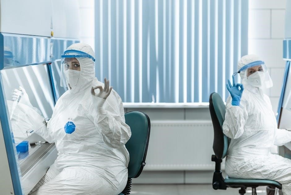
1.2 Proper Lab Attire and Personal Protective Equipment (PPE)
Appropriate attire is fundamental for safety in the biology lab. While specific requirements aren’t detailed in the provided text, general lab practice dictates protective measures. Closed-toe shoes are mandatory to shield feet from spills and broken glass. Clothing should minimize exposed skin, preventing contact with chemicals or biological materials.
Personal Protective Equipment (PPE), though not explicitly listed, is vital. This commonly includes safety goggles to protect eyes from splashes, and potentially gloves to prevent skin contact with substances. Lab coats offer an additional barrier, safeguarding clothing and skin.
Understanding and utilizing PPE correctly is crucial. Following instructor guidance on required equipment for each experiment ensures a safe and productive learning environment.
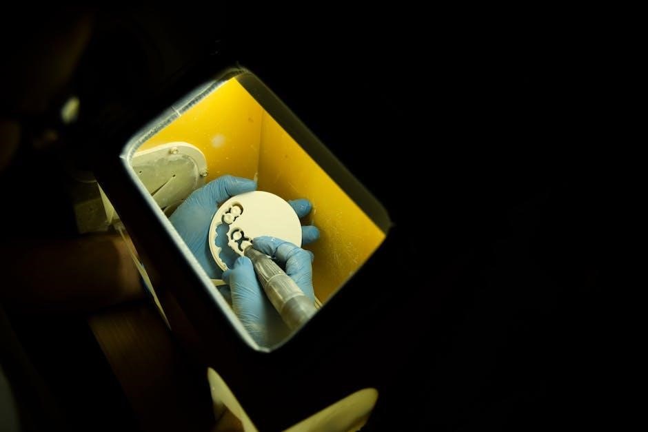
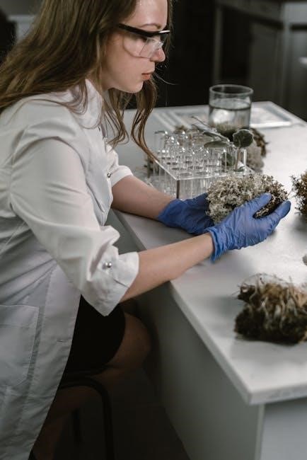
1.3 Handling and Disposal of Biological Materials
Safe handling of biological materials is paramount in the lab. While the provided excerpts don’t detail specific protocols, responsible practices are essential. Treat all specimens – even seemingly harmless ones – with respect and caution, assuming potential hazards. Avoid direct contact and follow instructor guidance meticulously.
Proper disposal procedures are critical to prevent contamination and ensure environmental safety. The manual doesn’t specify methods, but generally, biological waste requires designated containers separate from regular trash. Sharps, like slides or broken glass, demand specialized disposal to prevent injuries.
Adhering to these guidelines minimizes risks and maintains a safe laboratory environment for everyone. Always clarify disposal protocols with your instructor before proceeding.
Chapter 2: The Scientific Method and Experimental Design
This section focuses on the core principles of scientific investigation, including formulating hypotheses and designing controlled experiments for accurate data collection.
2.1 Formulating Hypotheses and Predictions
A crucial step in the scientific method involves developing testable hypotheses. These are proposed explanations for observed phenomena, acting as initial answers to specific questions. Hypotheses must be clear, concise, and falsifiable – meaning they can be proven wrong through experimentation.
Predictions logically follow from hypotheses, outlining expected outcomes if the hypothesis is true. For example, if a hypothesis states increased light enhances plant growth, a prediction might be: “Plants exposed to more light will exhibit greater biomass accumulation.”
Effective predictions are specific and measurable, allowing for objective evaluation of experimental results. This process ensures a rigorous and systematic approach to scientific inquiry, forming the foundation for reliable conclusions.
2.2 Identifying Independent and Dependent Variables
Experimental design hinges on correctly identifying variables. The independent variable is the factor deliberately manipulated by the researcher – it’s the presumed cause; For instance, in a plant growth study, light intensity could be the independent variable.
Conversely, the dependent variable is the factor measured to assess the effect of the independent variable – it’s the presumed effect. In the same study, plant height or biomass would be dependent variables.
Accurate identification is vital; the dependent variable’s changes are observed due to alterations in the independent variable. Control of other factors (constants) ensures only the independent variable influences the dependent variable, leading to valid conclusions.
2.3 Control Groups and Replication in Experiments
Robust experimental design necessitates both control groups and replication. A control group serves as a baseline for comparison, experiencing all conditions except the independent variable. This isolates the variable’s effect, demonstrating what happens without intervention.
Replication involves repeating the experiment multiple times with independent samples. This minimizes the impact of random errors and increases confidence in the results. Larger sample sizes enhance statistical power.
Without replication, observed effects might be due to chance, not the independent variable. Proper controls and sufficient replication are fundamental to drawing reliable, scientifically sound conclusions from biological investigations.

Chapter 3: Measurement and Data Analysis
Accurate measurement and data interpretation are vital in biology. This section covers the metric system, microscopy techniques, and effective data graphing methods.
3.1 The Metric System in Biology
The metric system is the standard for biological measurements, offering simplicity and ease of conversion. Utilizing units like meters, grams, and seconds ensures consistency and reduces errors in data collection. Lab work heavily relies on precise measurements, demanding familiarity with prefixes – kilo, centi, milli – to accurately represent quantities.
Understanding these prefixes allows for seamless conversion between units, crucial for calculations and data analysis. Consistent use of the metric system facilitates collaboration and reproducibility within the scientific community. Mastering this system is foundational for successful experimentation and interpretation of biological results, ensuring reliable and comparable data.
3.2 Using Microscopes and Measuring Techniques
Microscopes are fundamental tools in biology, enabling visualization of structures beyond the naked eye’s capability. Proper handling, including cleaning and focusing techniques, is essential for obtaining clear images. Lab exercises often involve measuring microscopic objects, requiring understanding of techniques like using ocular micrometers or stage micrometers.
Accurate measurements are crucial for characterizing cell sizes, identifying organelles, and quantifying biological features. Careful calibration of microscopes ensures reliable data. Mastering these skills allows for detailed observation and quantitative analysis, forming the basis for informed conclusions in biological investigations. Precise measurements contribute to the validity of experimental results.
3.3 Graphing and Interpreting Data
Data analysis is a cornerstone of the scientific method, and lab reports frequently require graphical representation of experimental results. Choosing the appropriate graph type – line, bar, or pie – depends on the nature of the data and the relationships being investigated. Accurate labeling of axes, including units, is paramount for clarity.
Interpreting graphs involves identifying trends, patterns, and anomalies. Students must be able to draw conclusions supported by the data, recognizing potential sources of error and limitations. Understanding statistical analysis, even at a basic level, enhances data interpretation and strengthens the validity of scientific claims.

Chapter 4: Biological Molecules
This section explores the fundamental building blocks of life: carbohydrates, lipids, and proteins. It also covers acids, bases, and the crucial role of pH in biological systems.
4.1 Structure and Function of Carbohydrates, Lipids, and Proteins
This lab exploration delves into the molecular world, examining the structures of carbohydrates – sugars and starches providing energy – lipids, like fats crucial for storage and insulation, and proteins, the workhorses of the cell.
Understanding their building blocks – monosaccharides, fatty acids, and amino acids respectively – is key. Experiments may involve testing for the presence of these molecules using specific reagents, revealing their unique chemical properties.
Focus will be placed on how these structures dictate function; for example, the folded shape of a protein determines its enzymatic activity. The manual guides students through identifying these molecules and relating their composition to their biological roles.
4.2 Acids, Bases, and pH in Biological Systems
This section of the manual focuses on the critical role of acids, bases, and pH in maintaining life’s processes. Experiments will likely involve using pH indicators to measure the acidity or alkalinity of various solutions, demonstrating the pH scale’s logarithmic nature.
Students will learn how biological systems, like blood, maintain a narrow pH range for optimal enzyme function and cellular activity. The lab may include buffering systems, exploring their ability to resist pH changes.
Understanding the concepts of hydrogen ion concentration ([H+]) and its impact on biological molecules is paramount. The manual provides guidance on safe handling of acids and bases, alongside interpreting experimental results.

Chapter 5: Cell Structure and Function
This lab explores plant and animal cells using microscopy, focusing on identifying key organelles and understanding their specific roles within cellular processes.
5.1 Observing Plant and Animal Cells Under a Microscope
This exercise introduces fundamental microscopy techniques for observing cellular structures; Students will prepare wet mount slides of both plant (like onion epidermis) and animal (cheek cells) tissues; Proper slide preparation is crucial, ensuring optimal visibility of cellular components.
Using the compound microscope, begin with the lowest power objective and gradually increase magnification. Focus carefully, noting the distinct differences between plant and animal cells. Observe the cell wall in plant cells, a structure absent in animal cells. Identify the nucleus, cytoplasm, and potentially other organelles depending on staining techniques.
Detailed observations and accurate drawings are essential for documenting findings. This hands-on experience builds a foundational understanding of cell morphology and prepares students for more advanced microscopic investigations.
5.2 Identifying Cell Organelles and Their Roles
Building upon microscopic observation, this section focuses on identifying key cell organelles and understanding their specific functions. Prepared slides of various cell types will be examined, often utilizing staining techniques to enhance visibility. Students will learn to recognize structures like the nucleus, mitochondria, ribosomes, and endoplasmic reticulum.
The nucleus, controlling center of the cell, and mitochondria, the powerhouses, are primary targets for identification. Understanding the role of each organelle in cellular processes – protein synthesis, energy production, waste removal – is paramount. Diagrams and illustrations will aid in correlating structure with function.
Accurate identification and a grasp of organelle roles are fundamental to comprehending cell biology and overall organismal function.
Chapter 6: Cell Transport Mechanisms
This chapter explores how substances move across cell membranes, focusing on diffusion, osmosis, and active transport. Experiments will demonstrate these principles, vital for cell function.
6.1 Diffusion and Osmosis: Principles and Experiments
Diffusion, the movement of molecules from high to low concentration, and osmosis, the diffusion of water across a semi-permeable membrane, are fundamental to cell life. This section details these processes, explaining how concentration gradients drive these movements.
Lab experiments will visually demonstrate diffusion using dyes and osmosis with plant cells in varying solutions. Students will observe how water potential affects cell turgor pressure, understanding the impact of hypotonic, hypertonic, and isotonic environments. Careful observation and data recording are essential for analyzing these transport mechanisms.
Understanding these principles is crucial for comprehending nutrient uptake, waste removal, and overall cellular homeostasis. The manual emphasizes safe handling of materials during these investigations.
6.2 Active Transport and Membrane Permeability
Active transport, unlike diffusion, requires energy to move molecules against their concentration gradient. This section explores mechanisms like protein pumps, vital for maintaining cellular imbalances. Membrane permeability dictates which substances can cross the cell membrane, influencing transport rates.
Lab activities may involve investigating the effects of inhibitors on active transport, demonstrating the energy dependence of these processes. Students will analyze how factors like temperature and molecule size affect membrane permeability. Precise measurements and controlled variables are key.
This manual stresses the importance of understanding how cells regulate their internal environment through these complex transport systems, ensuring proper function and survival.
Chapter 7: Enzyme Activity
This section details experiments investigating factors influencing enzyme catalysis, like temperature and pH. Students will explore enzyme specificity through controlled reactions and analysis.
7.1 Factors Affecting Enzyme Catalysis
Enzyme activity is profoundly influenced by environmental conditions. This experiment explores how variables like temperature and pH impact the rate of enzymatic reactions. Lab procedures will involve measuring reaction rates under differing conditions, observing how deviations from optimal levels diminish enzyme function.
Students will analyze data to determine the optimal temperature and pH for a specific enzyme. Understanding these factors is crucial, as enzymes operate within narrow ranges; extreme conditions can cause denaturation, permanently altering the enzyme’s structure and rendering it ineffective. Careful observation and precise measurements are essential for accurate results.
The manual emphasizes the importance of controlled experiments and replicates to ensure reliable conclusions about enzyme behavior.
7.2 Investigating Enzyme Specificity
Enzyme specificity, a cornerstone of biochemical reactions, dictates that each enzyme catalyzes only a specific substrate. This lab exercise focuses on demonstrating this principle through experimentation. Students will test an enzyme’s ability to react with various substrates, observing which combinations yield a measurable product.
The manual guides students through controlled setups, ensuring accurate observation of reaction outcomes. Data analysis will reveal which substrates are effectively processed by the enzyme, highlighting its selective nature. Understanding specificity is vital, as it underpins metabolic pathways and cellular regulation.
Precise measurements and careful documentation are crucial for validating the concept of enzyme-substrate interactions.

Chapter 8: Human Physiology – Cardiovascular System
This section of the manual details experiments measuring heart rate, blood pressure, and analyzing blood components, providing insights into cardiovascular function and health.
8.1 Heart Rate and Blood Pressure Measurements
This lab exercise focuses on accurately measuring vital signs – heart rate and blood pressure – to understand cardiovascular function. Students will learn proper techniques for pulse palpation and auscultation, utilizing stethoscopes for accurate heart sound detection.
Blood pressure measurement will involve using a sphygmomanometer, determining systolic and diastolic pressures. The manual emphasizes the importance of standardized procedures and recording data meticulously. Analyzing variations in heart rate and blood pressure under different conditions, like rest versus exercise, will be explored.
Understanding these measurements provides a baseline for assessing cardiovascular health and identifying potential physiological responses to stimuli. Safety protocols regarding equipment usage are highlighted throughout the procedure.
8.2 Analyzing the Components of Blood
This laboratory investigation delves into the composition of human blood, identifying its key components: plasma, red blood cells, white blood cells, and platelets. Students will utilize techniques like hematocrit determination to quantify red blood cell volume, gaining insight into oxygen-carrying capacity.
Microscopic examination of blood smears will allow for the identification and differentiation of various white blood cell types, crucial for immune function. The manual details staining procedures and proper slide preparation for optimal visualization.
Analyzing blood components provides a foundational understanding of its role in transport, immunity, and homeostasis. Safety precautions regarding handling blood samples and proper disposal are strictly enforced.

Chapter 9: Human Physiology – Respiratory System
This section explores lung capacity and ventilation rates, employing techniques detailed in the manual. Students will measure vital capacities, understanding breathing mechanics and gas exchange.
9.1 Lung Capacity and Ventilation Rates
This lab focuses on quantifying respiratory function, utilizing the manual’s procedures for measuring lung volumes and ventilation. Students will learn to determine vital capacity – the maximum air exhaled after maximal inhalation – using spirometers or simple water displacement methods.
Ventilation rate, the volume of air moved in and out of the lungs per minute, will also be assessed, both at rest and following exercise. The manual guides students through calculating these rates and analyzing the impact of physical activity on respiratory parameters.
Understanding these measurements provides insight into individual respiratory health and the physiological principles governing gas exchange, crucial for maintaining homeostasis.

Chapter 10: Dissection Techniques
This manual details frog dissection, emphasizing anatomical structures and functions. It provides step-by-step instructions and illustrations for safe, effective exploration of organ systems.
10.1 Frog Dissection: Anatomical Structures and Functions
Frog dissection, as outlined in this lab manual, offers a valuable hands-on experience in comparative anatomy. Students will systematically explore external features, then carefully dissect to reveal internal organ systems.
Key structures include the digestive system – stomach, intestines, liver, and pancreas – and the circulatory system, focusing on the heart and major blood vessels. The respiratory system, with lungs, and the urogenital system will also be examined.
Understanding the function of each structure is paramount. This exercise connects anatomy to physiology, illustrating how form dictates function. Precise identification and careful observation are essential for successful dissection and comprehension.
Leave a Reply
You must be logged in to post a comment.